It all began with an environmental catastrophe.
Roughly 1.5 to 2 billion years ago, Earth experienced what could only be described as a pollution crisis — oxygen levels surged dramatically.
Back then, most life on Earth was anaerobic. For these organisms, oxygen was a toxic gas that could wreak havoc on any life form relying on fermentation for energy.
Life, quite literally, teetered on the brink of extinction.
And just when things looked hopeless, a miraculous accident occurred:
A eukaryotic cell “swallowed” an alpha-proteobacterium. But instead of digesting it, the eukaryotic cell chose to… keep it. They lived together: the eukaryotic cell provided shelter and nutrients, while the engulfed bacterium offered a new, far more powerful energy mechanism — aerobic respiration1.
This ancient cohabitation led to one of the greatest energy revolutions in the history of life.
- Under anaerobic fermentation, a single glucose molecule yielded only 2 ATPs (adenosine triphosphate — life’s energy currency).
- But through aerobic respiration, the same glucose could generate 30–32 ATPs — a 16-fold leap in efficiency2,3.

Painted by AI
With more energy available, life could afford to become complex.
From multicellular organisms to warm-blooded animals — and eventually, to a particular primate species whose oversized, energy-hungry brain would usher in the age of Homo sapiens. The era of running, thinking, creating, and loving had begun.
That ancient bacterium? Today we call it mitochondria.
Not a parasite, but a partner
Our cells house hundreds of trillions of mitochondria4. And you can still see traces of their bacterial ancestry: they have their own DNA (mtDNA)5, their own ribosomes, and can synthesize a small set of proteins independently6. Their DNA still closely resembles that of free-living bacteria1.
Mitochondria are also inherited exclusively from the mother. That means your mitochondria can be traced back to a single ancestral woman hundreds of thousands of years ago — the so-called “Mitochondrial Eve.”
In the cult sci-fi horror novel Parasite Eve, mitochondria evolve consciousness and attempt to take over their human hosts to fulfill their own evolutionary goals. The book sold over 1.5 million copies in Japan and inspired a globally popular video game.

Image from internet
But one thing is certain: mitochondria are not parasites. They’ve formed a truly mutualistic symbiosis with their host. The host provide them with shelter and nutrients; in return, they deliver a steady, efficient supply of energy.
Over time, mitochondria and their host have become so deeply integrated that they’re now inseparable — no longer capable of surviving on their own.
So no, you don’t need to worry about your mitochondria “rebelling” like in those sci-fi novels or video games.
Mitochondrial efficiency is a core marker of Prime Life
Roughly 90% of our body’s energy is produced by mitochondria. A 70-kg adult generates about 70 kilograms of ATP per day via mitochondrial respiration7,8.
As a PNAS commentary once put it: “In a single day, the human body cycles through its entire bodyweight in ATP.”
Some organs rely on mitochondria more than others. Cardiac muscle cells can house up to 5,000 mitochondria each, taking up nearly one-third of the cell’s volume. Neurons, too, are packed with tiny energy plants9,10.
And we all know what it feels like when mitochondria are working well:
- That post-workout high, the clarity of thought — it’s your mitochondria in overdrive.
- And when you feel tired, sluggish, foggy, slow to recover — it’s not necessarily “kidney deficiency” (as some traditional ideas suggest). More likely, it’s mitochondrial inefficiency.
In 2013, the journal Cell named “mitochondrial dysfunction” as one of the primary hallmarks of aging. As we grow older, mtDNA accumulates damage, biosynthetic efficiency drops, oxidative stress builds up. In plain terms: the “power plants” fueling your body keep breaking down, and everything else starts to follow.
In a way, aging is the story of mitochondria shutting down.
That’s why mitochondrial function is central to both our Health Span and our daily vitality — and why it’s a core metric of a Prime Life.
Exercise is the best way to boost mitochondrial function
There are many ways to stimulate mitochondria through mild stress — exercise, caloric restriction, fasting, cold/heat exposure, intermittent hypoxia… even supplements like NAD+ (or precursors like NR/NMN), and CoQ10.
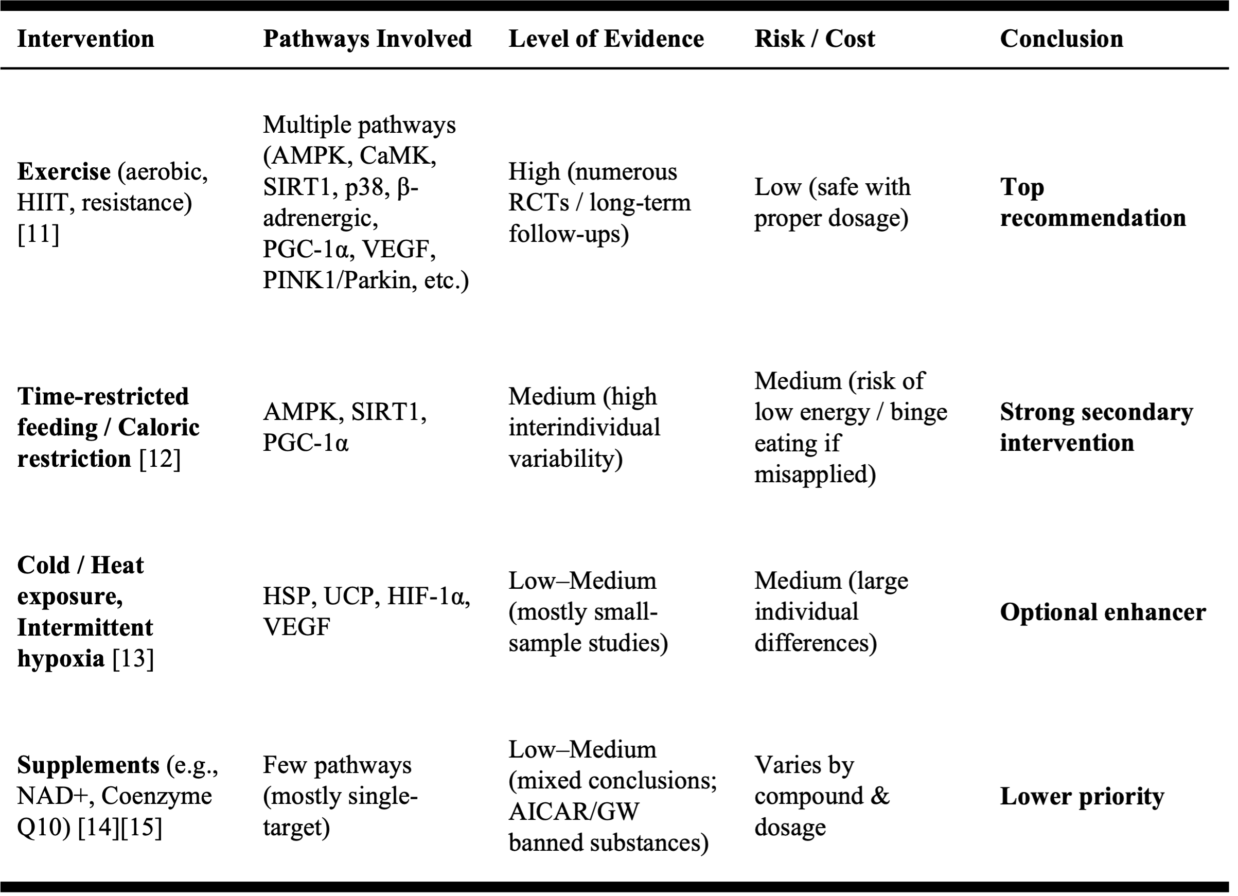
But among all these, exercise stands alone as the single most effective intervention16:
- Numerous RCTs and long-term cohort studies support exercise’s benefit on mitochondrial health.
- It activates multiple pathways: AMPK/PGC-1α, mitochondrial biogenesis, autophagy, antioxidant defense.
- It’s safe, evolutionarily aligned, systemic, and sustainable.
- And its cost is essentially zero — all you need is a pair of shoes.
The ROI of exercise is nearly infinite:
minimal input, maximal return.
The AMPK–PGC-1α Axis: Exercise’s Mitochondrial Pathway
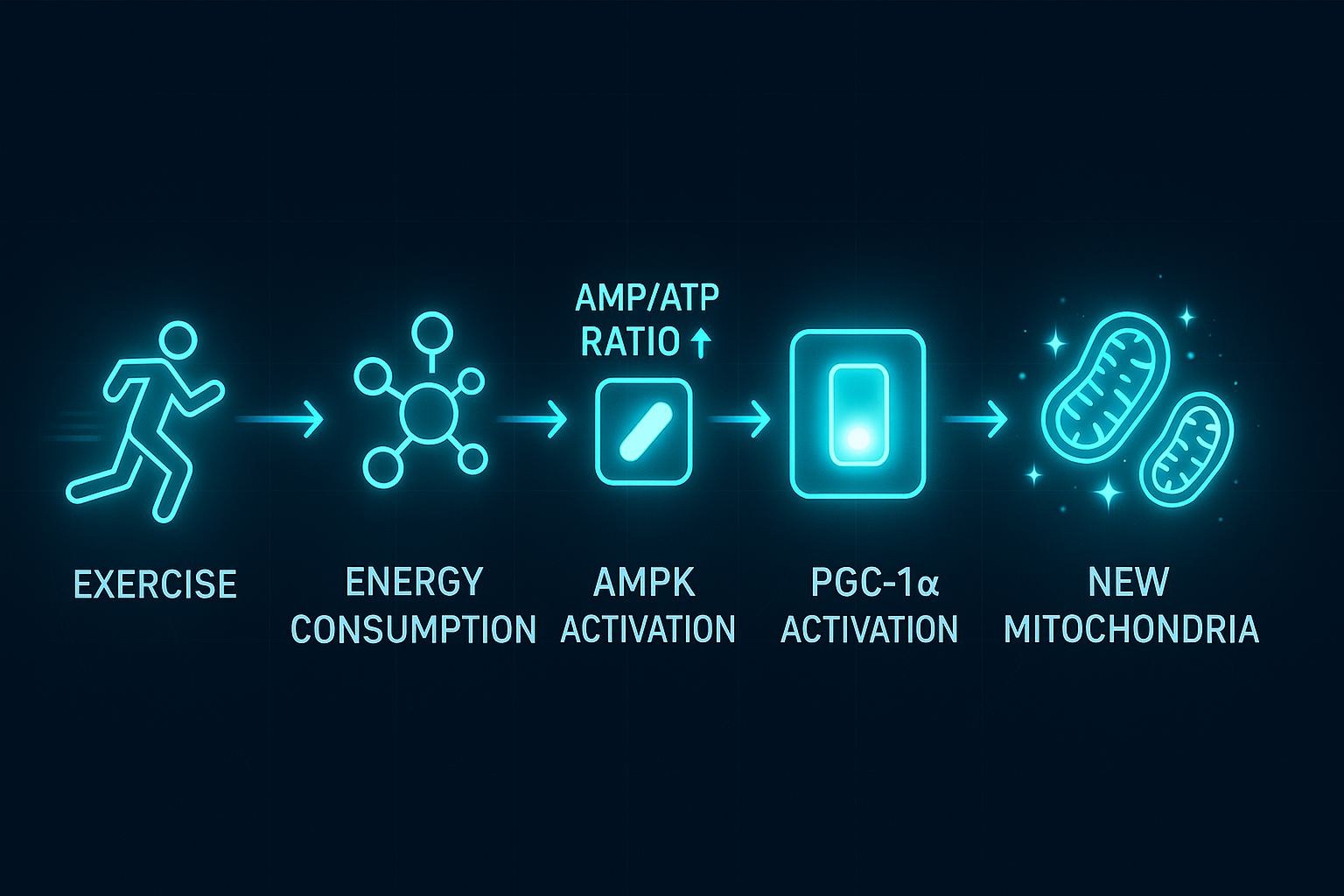
Exercise → Energy expenditure → Increased AMP/ATP ratio → AMPK activation.
AMPK then activates PGC-1α, the master switch that recruits downstream factors like NRF-1 and TFAM, launching mitochondrial biogenesis17.
- Moderate-intensity exercise (60–70% VO₂max): AMPK activation increases 3–5x18
- High-intensity exercise (>85% VO₂max): AMPK activation can increase up to 10x19
The result? More mitochondria, stronger function, better endurance, greater metabolic flexibility.
Painted by AI
Why do we feel amazing after exercise?
Unlike other interventions, exercise offers something unmatched: instant feedback, crystal-clear body sensations.
Why do you feel invigorated after a workout?
Because you’ve just activated the acute effects of mitochondrial upregulation. ATP demand increases 100 to 1000 times, mitochondrial ATP and oxygen throughput jumps 5–10 fold during exercise20,21.
Why does your thinking feel sharper?
Because just 30 minutes of movement boosts BDNF (brain-derived neurotrophic factor) levels by 200–300% — think of it as brain fertilizer[22–24]. Combined with increased ATP delivery, your cognitive clarity spikes.
Why do your steps feel lighter?
Because post-exercise, your insulin sensitivity improves, glucose uptake increases, glycogen replenishment accelerates, and your energy turnover is smoother. At the same time, skeletal muscle and cardiac muscle preferentially oxidize lactate, helping restore metabolic balance faster.
These are short-term fluxes — the immediate effects.
But over days to weeks, cumulative training leads to lasting mitochondrial gains: increased content, improved respiration, better endurance, more efficient fuel switching (fat ↔ glucose), and a smoother, more energetic baseline for everyday life[25–28].
This is why exercise is the #1 method for activating and maintaining mitochondrial health.
In Summary
Starting our conversation on exercise by diving into mitochondria serves two purposes:
It explains a core aging mechanism, and it gives us a foundational reason to commit to movement as a tool for health and longevity.
Mitochondria represent one of the most extraordinary symbiotic relationships in evolutionary history. A swallowed bacterium changed the fate of life on Earth — enabling complexity, consciousness, and, ultimately, us.
Today, they still hum away inside us — powering every thought, heartbeat, and step.
They made us human. And when they falter, we age.
Exercise is how we honor that ancient pact. Not pills. Not hacks. Just movement.
In our pursuit of a Prime Life, there are countless longevity strategies. But sometimes the simplest is also the most powerful — move.
Because let’s face it:
Doomscrolling won’t build mitochondria. But running will.

Painted by AI
Intervention recommendations are geared toward generally healthy individuals. If you have existing health conditions, always follow your doctor’s instructions.
References28
[1]Gray, Michael W. “Mitochondrial Evolution.” Cold Spring Harbor Perspectives in Biology, vol. 4, no. 9, 2012, a011403.
[2]Hinkle, Peter C. “P/O Ratios of Mitochondrial Oxidative Phosphorylation.” Biochimica et Biophysica Acta (Bioenergetics), vol. 1706, 2005, pp. 1–11.
[3]Chaudhry, Rida, and Richard T. Ellison. “Biochemistry, Glycolysis.” StatPearls, StatPearls Publishing, 2023.
[4]BioNumbers. “How Big Are Mitochondria?” BioNumbers Book, Harvard Medical School, accessed 8 Sept. 2025.
[5]Taanman, Jan-Willem. “The Mitochondrial Genome: Structure, Transcription, Translation and Replication.” Biochimica et Biophysica Acta, vol. 1410, no. 2, 1999, pp. 103–123.
[6]Greber, Björn J., et al. “The Complete Structure of the 55S Mammalian Mitochondrial Ribosome.” Science, vol. 348, no. 6232, 2015, pp. 303–308.
[7]Törnroth-Horsefield, Susanna, and Richard Neutze. “Opening and Closing the Metabolite Gate.” Proceedings of the National Academy of Sciences, vol. 105, no. 50, 2008, pp. 19565–66.
[8]BioNumbers. “Daily Turnover of ATP — Human (BNID 105606).” Harvard Medical School, accessed 8 Sept. 2025.
[9]Li, A., et al. “Mitochondrial Dynamics in the Adult Cardiomyocytes and Cardiac Diseases.” Frontiers in Cell and Developmental Biology, 2020, Article 584800.
[10]Wang, Xin, et al. “Mitochondrial Flashes Regulate ATP Homeostasis in the Heart.” eLife, 2017, e23908.
[11]MacInnis, Martin J., and Martin J. Gibala. “Physiological Adaptations to Interval Training and the Role of Exercise Intensity.” Physiological Reviews, 2017.
[12]de Cabo, Rafael, and Mark P. Mattson. “Effects of Intermittent Fasting on Health, Aging, and Disease.” NEJM, 2019.
[13]Cypess, Aaron M., et al. “Identification and Importance of Brown Adipose Tissue in Adult Humans.” NEJM, 2009.
[14]Dollerup, Ole L., et al. “Nicotinamide Riboside Does Not Alter Mitochondrial Function or Body Composition in Obese Men.” JCI Insight, 2018.
[15]Yoshino, Jun, et al. “Nicotinamide Mononucleotide Increases Muscle Insulin Sensitivity in Prediabetic Women.” Science, 2021.
[16]Bishop, David J. “Exercise as Mitochondrial Medicine.” Annual Review of Physiology, 2025; Mølmen, Kristoffer S., et al. “Effects of Exercise Training on Mitochondrial and Capillary Content.” Sports Medicine, 2025.
[17]Jäger, Sebastian, et al. “AMP-Activated Protein Kinase (AMPK) Action in Skeletal Muscle via Direct Phosphorylation of PGC-1α.” Proceedings of the National Academy of Sciences, vol. 104, no. 29, 2007, pp. 12017–22.
[18]Stephens, T. J., et al. “Progressive Increase in Human Skeletal Muscle AMPKα2 Activity and ACC Phosphorylation during Exercise.” American Journal of Physiology–Endocrinology and Metabolism, vol. 282, no. 3, 2002, pp. E688–E694.
[19]McConell, Glenn K., et al. “Letter to the Editor: Skeletal Muscle AMPK Is Activated during Prolonged Exercise in Humans.” J Physiol, 2020.
[20]Baker, Julien S., et al. “Metabolic Response to High Intensity Exercise.” Sports Medicine, 2010.
[21]Boushel, Robert, et al. “Maximal Oxygen Uptake …” Experimental Physiology, 2011
[22]Szuhany, Kristin L., et al. “A Meta-Analytic Review of the Effects of Exercise on BDNF Levels.” J Psychiatr Res, 2015.
[23]Dinoff, Alex, et al. “The Effect of Acute Exercise on Circulating BDNF in Humans: A Meta-Analysis.” Neuroscience Letters, 2017
[24]Rasmussen, Peter, et al. “Evidence for a Release of Brain-Derived Neurotrophic Factor from the Brain during Exercise.” J Physiol, 2009.
[25]Mikines, K. J., et al. “Enhanced Insulin Action on Skeletal Muscle Glucose Transport and Uptake After a Single Bout of Exercise.” The Journal of Clinical Investigation, 1988.
[26]Brooks, George A. “The Science and Translation of Lactate Shuttle Theory.” Cell Metabolism, 2018.
[27]Bishop, David J. “Exercise as Mitochondrial Medicine: How Does the Exercise Prescription Affect Mitochondrial Adaptations to Training?” Annual Review of Physiology, 2025.
[28]Goodpaster, Bret H., and L. M. Sparks. “Metabolic Flexibility in Health and Disease.” Cell Metabolism, 2017.